

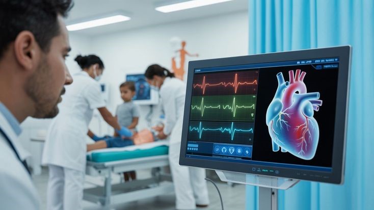
The diagnostic path for a heart condition often begins with a familiar, non-invasive experience. For forty-five-year-old Nitin, an operations head at an engineering firm in Chinchwad, persistent breathlessness during his evening walks led him to a cardiac consultation. In the clinic’s evaluation room, the medical team applied a cool gel to his chest and glided a smooth plastic probe across his ribs, displaying a real-time, moving black-and-white image of his heart on a monitor.
This test, a 2D Echocardiogram (Echo), is the universal workhorse of cardiac diagnostics. It quickly showed that Nitin’s heart muscle was abnormally thick and struggling to relax—clear indicators of a cardiomyopathy.
However, instead of finalizing a treatment plan right then, the cardiologist looked at the ultrasound loops and delivered an unexpected recommendation: “Nitin, the Echo tells us your heart muscle is thick, but to find out exactly why it is thick, we need to schedule a Cardiac MRI.”
Nitin was understandably confused. Why undergo a second, more complex scan inside a loud, metallic tube when a high-tech ultrasound machine had already looked directly at his beating heart?
For many patients navigating a cardiomyopathy or heart muscle disease, understanding the diagnostic pipeline is key to peace of mind. Cardiologists don’t deploy these tests randomly. The relationship between a 2D Echo and a Cardiac Magnetic Resonance (CMR) scan is a structured, complementary partnership—moving from rapid structural mapping to microscopic tissue analysis.
The First Responder: Why the 2D Echo Always Comes First
A cardiac evaluation virtually always begins with a 2D Echocardiogram. An Echo uses high-frequency sound waves to construct real-time moving images of the heart’s architecture. It is the definitive “first responder” of cardiac diagnostics for several practical and clinical reasons:
- Instantaneous, Real-Time Feedback: An Echo allows a cardiologist to watch your heart valves open and shut dynamically and observe the synchronized squeeze of your muscle walls beat by beat.
- Accessibility and Comfort: The test is completely non-invasive, radiation-free, requires no advance preparation, and can be completed at the bedside in less than twenty minutes.
- The Baseline Blueprint: An Echo provides immediate answers to foundational structural questions. It measures the precise thickness of your heart walls, maps the size of your internal chambers, and calculates your ejection fraction (the percentage of blood pumped out with each contraction).
For a vast number of routine cardiac issues—like assessing standard valve wear or checking overall pumping strength after a mild heart attack—a high-resolution 2D Echo provides all the data a clinician needs to guide therapy.
The Specialist: When a Cardiologist Deploys Cardiac MRI
If a 2D Echo is so efficient, why look further? The answer lies in a fundamental limitation of ultrasound technology. Sound waves are excellent at mapping the shape, motion, and dimensions of a structure, but they cannot look through the tissue to analyze its internal cellular composition. An Echo can tell a doctor that a heart wall is thick or weak, but it cannot easily differentiate whether that weakness is caused by generic scar tissue, abnormal protein infiltration, or unusual iron deposits.
This is exactly where Cardiac MRI (CMR) becomes indispensable. A Cardiac MRI utilizes powerful magnetic fields and radio waves to generate ultra-high-resolution, three-dimensional images of the heart. Its true superpower is tissue characterization—the ability to identify the exact health, makeup, and cellular environment of the heart muscle fibers.
Cardiologists transition from an Echo to an advanced Cardiac MRI under specific clinical scenarios:
- Differentiating Complex Cardiomyopathies
If an Echo reveals a severely thickened heart wall, a Cardiac MRI is deployed to pinpoint the root cause. By analyzing the way magnetic signals bounce off the tissue, a CMR can distinguish between Hypertrophic Cardiomyopathy (HCM) (a genetic muscle disarray), Cardiac Amyloidosis (the infiltration of misfolded proteins), or Fabry disease (a rare metabolic storage disorder). Each of these conditions looks similar on a basic ultrasound but requires vastly different medical treatments.
- Mapping Scar Tissue (Late Gadolinium Enhancement)
During a Cardiac MRI, a specialized, safe contrast agent called gadolinium is injected into the bloodstream. Healthy, tightly packed heart muscle cells flush the contrast dye out rapidly. However, areas of the heart that are scarred, dead, or inflamed retain the contrast agent, lighting up brightly on the scan.
This technique, known as Late Gadolinium Enhancement (LGE), allows cardiologists to see the exact location, depth, and pattern of scar tissue within the living heart wall. Mapping this scar pattern helps determine if a patient’s muscle weakness stems from a past silent heart attack (ischemic) or an active inflammatory muscle disease like Myocarditis or Sarcoidosis (non-ischemic).
- Resolving Poor Acoustic Windows
Ultrasound sound waves must travel through skin, fat, muscle, and bone to reach the heart. In patients with specific body types, severe lung disease (like emphysema), or thick chest walls, the Echo images can occasionally turn out blurry or inconclusive. Because an MRI uses magnetic fields rather than sound waves, it easily bypasses these physical barriers, providing pristine, sub-millimeter visual clarity regardless of a patient’s body habitus.
Defining the Diagnostic Shift
While a 2D Echocardiogram excels as a first-line structural screening tool—providing rapid, silent, and real-time feedback on valve motion and basic pumping volume—the Cardiac MRI functions as an advanced, second-line problem solver. The primary difference lies in how they evaluate tissue. An Echo treats the heart walls as solid, moving architectural structures, whereas a Cardiac MRI delivers exceptional cellular detail, identifying hidden fat, iron, protein deposits, and microscopic scars.
The initial ultrasound provides the baseline blueprint in about fifteen minutes right in the clinic room, while the longer MRI scan requires a specialized magnet tunnel to unlock the precise biological answers hidden deep within the muscle fibers.
Advanced Diagnostic Precision at Dr. Akshay Kashid’s Clinic
Navigating a progressive heart muscle disorder requires moving past generalized checkups and embracing highly precise, data-driven imaging pathways. Ensuring long-term cardiovascular health depends on choosing the correct diagnostic tools at the right clinical moments to build an accurate, personalized roadmap for your heart.
At the Dr. Akshay Kashid Heart Care Clinic in Balewadi-Baner, Pune, a dedicated structural diagnostic framework guides patients seamlessly through their clinical evaluations:
- Definitive First-Line Imaging (Advanced 2D Echo): For an immediate, high-resolution look at your heart’s primary architecture, Dr. Kashid utilizes precise 2D Echo Test In Pune diagnostics. This essential ultrasound screening allows the clinical team to instantly track your heart chamber volumes, check valve integrity, and calculate your baseline pumping metrics under comfortable, in-clinic conditions.
- Electrical Network Profiling: Structural muscle shifts often leave distinct, telltale markers on your heart’s electrical grid. The clinic provides comprehensive ECG Test In Pune assessments and ambulatory rhythm tracking to capture conduction delays or silent rhythm irregularities early.
- Functional Workload Surveillance: To see exactly how your heart muscle and blood pressure behave when subjected to increasing workloads, Dr. Kashid integrates a structured, treadmill-based Stress Test In Pune. This safely exposes any underlying drops in exercise tolerance or hidden circulation limitations under stress.
- Systemic Vascular and Plaque Controls: Protecting a compromised heart muscle involves minimizing any extra resistance in your arteries. Dr. Kashid coordinates specialized High Blood Pressure Treatment In Pune and advanced Cholesterol Treatment In Pune to stabilize your blood vessels and control systemic inflammation, creating a stable environment for your heart to recover.
- Expert Advanced Imaging Referral & Insight: When a baseline Echo points to a complex tissue disorder, a Cardiac MRI is carefully coordinated. As an accomplished interventional cardiologist practicing at premier regional medical institutions, Dr. Akshay Kashid personally interprets complex CMR tissue reports and scar maps, translating intricate radiological data into clear, actionable treatment plans for families.
Conclusion: Layering Technology for Ultimate Clarity
A 2D Echo and a Cardiac MRI should never be viewed as competing tests; they are successive steps on a pathway designed to provide absolute clinical certainty. The human heart is a complex, multi-layered machine, and managing its conditions effectively requires understanding both its macroscopic movements and its microscopic tissue health. By utilizing a first-line ultrasound to map the foundation and deploying advanced magnetic resonance imaging when tissue details are needed, your medical team can remove all guesswork—ensuring your heart receives the precise, targeted care it needs to thrive safely for the long term.
If you are experiencing unexplained shortness of breath, unusual fatigue, or have been told you have an enlarged heart or an abnormal heart murmur, take a proactive step toward clarity today.
To establish your structural cardiac baseline or discuss an advanced imaging pathway, schedule an expert consultation at the Dr. Akshay Kashid Heart Care Clinic. Reach out directly to our dedicated clinical team by calling 07058362823, or visit us in person at Office no. 301, 3rd Floor, V Business Center, S. no. 9/16/1 & 2, Near Lakshmi-Mata Mandir, Balewadi-Baner, Pune, Maharashtra 411045. Ensure your heart health is guided by a clear, uncompromised view