The persistent, dry evening cough had been bothering thirty-eight-year-old Sunita for over four months. Living in a bustling residential area of PCMC, she assumed it was just a lingering reaction to urban dust or an extended post-viral allergy. But lately, her daily routine felt increasingly heavy. Simply walking up the gentle incline to her local vegetable market left her heart pounding violently against her ribs, her breath coming in shallow, desperate gasps.
When she mentioned a history of severe, untreated throat infections during her childhood, the diagnostic path shifted instantly.
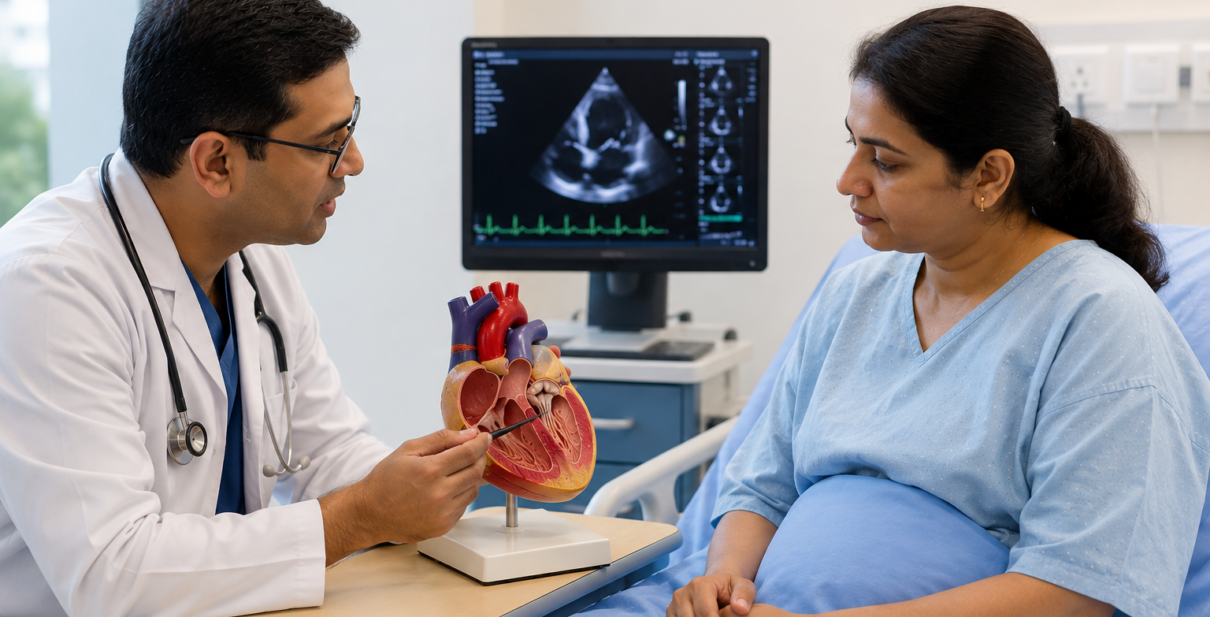
A specialized cardiac ultrasound revealed a troubling reality: Sunita’s heart was struggling against a decades-old echo of her youth. A condition known as Rheumatic Heart Disease (RHD) had left her primary inflow valve heavily scarred, thickened, and fused shut. Her heart was working under intense, unnatural strain just to push blood through a tiny, restricted opening.
For many young and middle-aged adults across Pune, managing the progressive exhaustion of Rheumatic Mitral Stenosis can feel like a losing battle fought with endless pill strips and water-retention tablets. But when a scarred heart valve narrows past a critical threshold, medication alone can no longer clear the bottleneck. This is exactly where an advanced, non-surgical structural intervention known as Balloon Mitral Valvotomy (BMV) becomes a life-changing turning point.
The Silent Attack: How Rheumatic Fever Scars the Mitral Valve
To understand why a structural intervention becomes necessary, we have to trace the disease back to its origin. Rheumatic Heart Disease is the long-term consequence of an unmanaged childhood bout of rheumatic fever, which often starts as a simple streptococcal sore throat. If the initial infection goes untreated, the body’s immune system can become confused, launching a mistaken attack against its own tissues—specifically targeting the delicate architecture of the Mitral Valve.
The mitral valve serves as a critical internal gateway, positioned between the heart’s upper left holding chamber (the left atrium) and its primary lower pumping chamber (the left ventricle). A healthy mitral valve consists of two flexible, paper-thin tissue leaflets that open wide with every heartbeat, allowing a smooth, rapid flow of oxygen-rich blood to fill the main pump.
Over the ten to twenty years following a rheumatic fever event, a slow process of chronic inflammation takes place within the heart:
- Commissural Fusion: The delicate edges where the valve leaflets meet (the commissures) begin to scar, thicken, and physically fuse together.
- The Narrowing Gateway (Mitral Stenosis): The once-wide valve opening progressively shrinks, transforming from a spacious gateway into a rigid, narrow slit.
- The Back-Pressure Cascade: Because blood cannot flow easily into the main pumping chamber, fluid backs up into the left atrium and the delicate blood vessels of the lungs. This chronic back-pressure is what causes patients to experience persistent breathlessness, nighttime coughing fits, and fluid retention.
The Non-Surgical Breakthrough: How BMV Restores Flow
For decades, the only definitive solution for a severely blocked mitral valve was complex open-heart surgery, requiring doctors to split the breastbone, stop the heart entirely, and manually cut open or replace the damaged valve. However, advanced interventional cardiology has introduced a remarkably elegant, minimally invasive alternative: Balloon Mitral Valvotomy (BMV), also frequently referred to as Percutaneous Transvenous Mitral Commissurotomy (PTMC).
Instead of making large incisions in the chest, a skilled interventional cardiologist can repair the structural defect entirely from the inside out using the body’s natural vascular highways:
- Vascular Access: Under local anesthesia, a thin, flexible hollow tube called a catheter is inserted into a major vein in the patient’s groin (the femoral vein) and gently guided upward into the heart.
- Crossing the Septum: The catheter carefully crosses the thin muscular wall dividing the right and left upper chambers of the heart, granting the specialist direct access to the narrowed mitral valve.
- The Precision Inflation: A specialized, high-pressure balloon (commonly an Inoue balloon) is guided directly across the rigid, fused opening of the mitral valve. Once perfectly positioned under real-time X-ray imaging, the balloon is rapidly inflated for just a few seconds.
The physical expansion of the balloon exerts a powerful, radial outward force. This precisely tears apart the scarred, fused edges of the leaflets along their natural lines of separation, instantly widening the valve opening and restoring a healthy, unrestricted pathway for blood flow without touching a single bone or muscle in the chest.
When is BMV the Right Clinical Choice?
While BMV is an incredibly effective procedure, it is not a universal fix for every individual with mitral valve disease. Determining whether a patient is a suitable candidate requires a highly detailed anatomical evaluation using a specialized scoring system (such as the Wilkins Score) to assess the valve’s unique structure.
Ideal Candidates for BMV
- High Valve Flexibility: The structural leaflets must remain relatively mobile and free from massive, dense calcium deposits that would prevent a balloon from opening the pathways cleanly.
- Minimal Sub-Valvular Thickening: The complex web of tendons and muscles beneath the valve should not be severely fused or shortened.
- Absence of Severe Leaking: The patient must not have significant pre-existing mitral regurgitation (a severe backward leak), as balloon expansion can potentially worsen a leaking valve.
- No Left Atrial Clots: A comprehensive ultrasound must confirm that no blood clots have formed in the upper chamber due to stagnant blood flow, ensuring a safe pathway for the procedure.
Specialized Structural Care at Dr. Akshay Kashid’s Clinic
Deciding on the right path for a progressive heart valve condition requires a shift away from temporary solutions and a reliance on advanced clinical data. Navigating a condition like Rheumatic Heart Disease demands an experienced medical hand capable of evaluating both the electrical and physical structures of the heart.
At the Dr. Akshay Kashid Heart Care Clinic in Balewadi-Baner, Pune, a comprehensive clinical and diagnostic pathway ensures patients receive optimal structural care:
- Definitive Visual Diagnostics (Advanced 2D Echo): The vital first step in determining your suitability for a balloon procedure is a high-resolution 2D Echo Test In Pune. Dr. Kashid utilizes precise cardiac ultrasound imaging to measure the exact surface area of your valve opening, calculate pressure gradients, and meticulously score your valve structure to ensure a safe, successful outcome.
- Vascular Stress Profiling: To verify how your heart muscle and lungs are coping with the narrowed gateway, the clinic utilizes targeted diagnostics, including a professional Stress Test In Pune and preventative ECG Test In Pune screenings to monitor electrical rhythms and rule out concurrent issues like atrial fibrillation.
- Systemic Management and Aftercare: Managing a structural valve condition involves protecting the entire circulatory system. Dr. Kashid integrates specialized High Blood Pressure Treatment In Pune and targeted Cholesterol Treatment In Pune to minimize any extra resistance in your arteries, helping your heart operate as smoothly as possible before and after an intervention.
- Experienced Interventional Oversight: As a highly trained interventional cardiologist with an extensive background performing complex catheter-based therapies at leading medical centers, Dr. Akshay Kashid personally guides patients through the entire procedural pipeline—from initial diagnostic evaluation to hospital-based interventional execution and long-term recovery.
Conclusion: Opening the Pathway to a Vibrant Life
Your breathing shouldn’t feel like an exhausting daily battle against a scarred and restricted heart valve. The human heart is a remarkably resilient pump, but when a structural bottleneck like mitral stenosis takes hold, it requires a definitive, mechanical solution to restore its natural rhythm. Choosing to address a narrowing valve through a modern, non-surgical procedure like a Balloon Mitral Valvotomy allows you to clear the obstruction safely, giving your heart the space and freedom it needs to power your life without restrictions.
If you or a loved one are managing the progressive breathlessness of Rheumatic Heart Disease, or if a routine checkup has revealed a narrowed mitral valve, don’t wait for your symptoms to compromise your independence.
To evaluate your structural cardiac baseline or discuss advanced therapeutic options, schedule a clinical consultation at the Dr. Akshay Kashid Heart Care Clinic. Reach out to our dedicated team directly by calling 07058362823, or visit us in person at Office no. 301, 3rd Floor, V Business Center, S. no. 9/16/1 & 2, Near Lakshmi-Mata Mandir, Balewadi-Baner, Pune, Maharashtra 411045. Give your heart the clear, open pathway it deserves.